Areas w/ no BBB - choroid plexus or pituitary or pineal gland or tuber cinereum (controls circadian rhythm in the inferior hypothalamus) or area postrema (vomiting in the inferior aspect of the fourth ventricle)
Vascular enhancement is localized increase of blood flow - caused by vasodilation or hyperemia or neovascularity or arteriovenous shunting
Periventricular enhancement can be neoplastic or infectious or demyelinating or treatment (radiation) induced
Nodular subcortical enhancement is usually metastatic disease (usually accompanied by edema). If disease is localized to posterior fossa might be venous spread of pelvic malignancy.
Gyriform enhancement - superficial enhancement due to infection or inflammation or ischemia (differential includes herpes encephelitis or meningitis or subacute infarct or PRES (posterior reversible encephalopathy syndrome) or SMART (stroke like migraine attacks after radiation therapy).
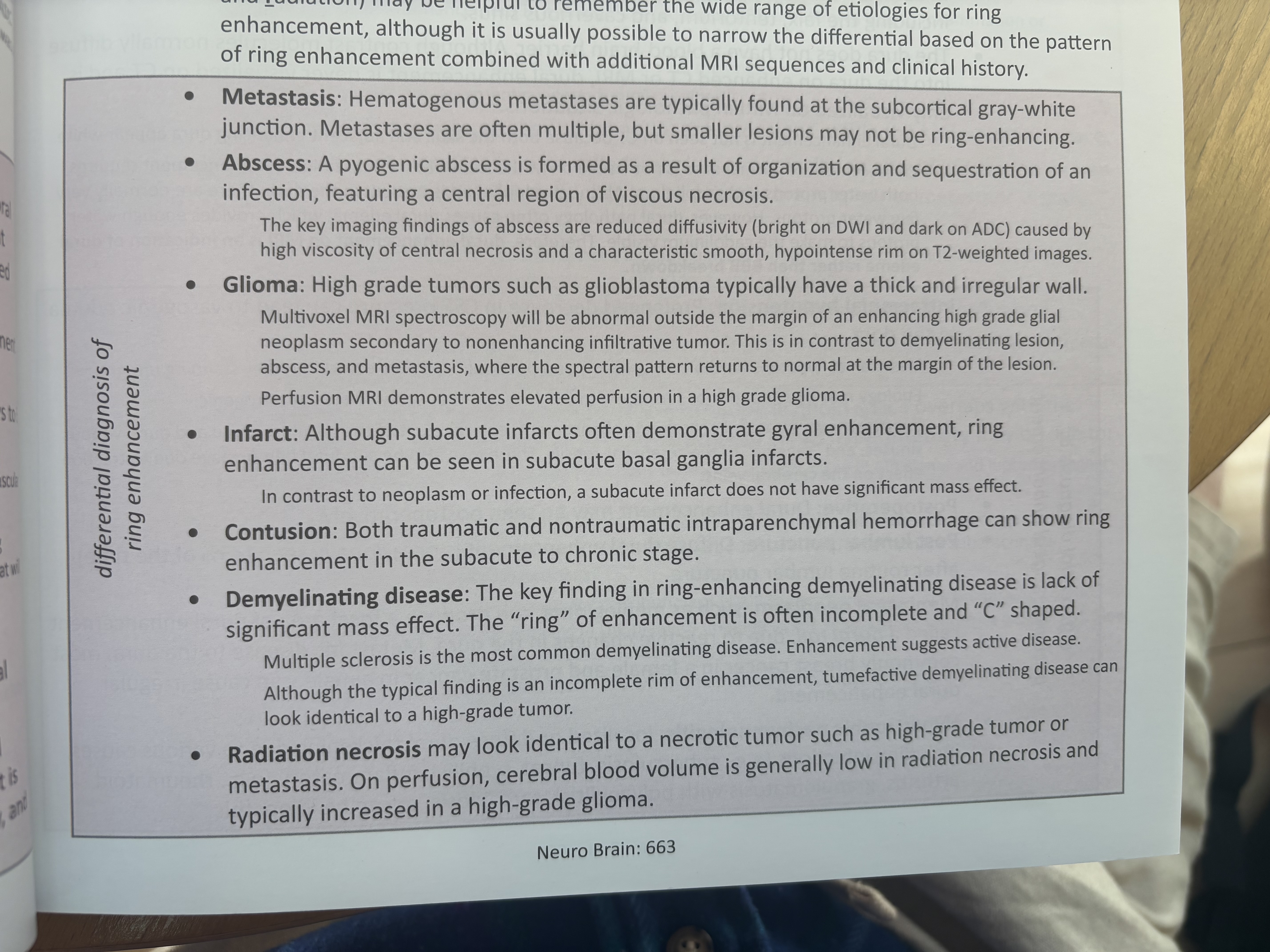
Ring enhancement (mnemonic MAGIC DR - metastasis abscess glioma infarct contusion demylenation radiation).
Pachymeningeal Enhancement - Only ever visualized on MR. Usually there isn't water in the dura so virual al dural enhancement on MRI is indication of dural edema rather than BBB breakdown. Sources include intracranial hypotension (leading to edema) OR post op OR post LP OR miningeal neoplasm OR hypertrophic pachymeningitis (which is localized due to infectious process like TB or funal or syphilis or inflammatory like RA or granulomatosis w/ polyangiitis or sarcoidosis) or idiopathic.
Leptomeningeal (pia-arachnoid) enchancement (extra axial) - caused by meningitis or encephalitis or metastases or amlyoidosis. Be careful to also differentiate w/ subarachnoid FLAIR hyperintensity differential which includes the above infectious and metastatic processes plus subarach hemorrhage and slow vascular flow (pt on propofol).